Dupuytren's Disease — TGF-β1 Protein Inhibitor Design
Research ownership & attribution: Rami M. Alkhaleeli — Alkhaleeli BioAI LLC
Scientific Concept
Dupuytren's disease is driven by TGF-β1 signaling, which promotes fibroblast activation, myofibroblast differentiation and extracellular matrix/collagen deposition in fibrotic palmar tissue. The molecular target is TGF-β1 itself. This computational target analysis maps the TGF-β1 precursor domains — a 29-aa signal peptide, a 249-aa latency-associated peptide (LAP) and a 112-aa mature domain (~408 aa total) — and designs a high-affinity soluble inhibitor (e.g., a LAP variant or receptor trap) intended to neutralize fibrotic TGF-β1 signaling, encoded in mRNA for local expression at the diseased tissue site.
Scientific Diagram
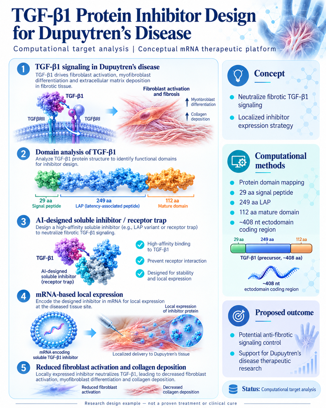
Original uploaded scientific infographic — use the Enlarge button for a full-size view.
Design Process
- 1
Signaling analysis — map how TGF-β1 drives fibroblast activation, myofibroblast differentiation and collagen deposition via TGFβRI/II.
- 2
Domain analysis of TGF-β1 — analyze the protein structure to identify functional domains: 29-aa signal peptide, 249-aa LAP, 112-aa mature domain.
- 3
AI-designed soluble inhibitor / receptor trap — design a high-affinity soluble inhibitor to neutralize fibrotic TGF-β1 signaling and prevent receptor interaction.
- 4
mRNA-based local expression — encode the designed inhibitor in mRNA for local expression at the diseased tissue site (ectodomain coding region ~408 nt).
- 5
Fibrosis-impact modeling — model reduced fibroblast activation and decreased collagen deposition.
Proposed Biological Mechanism
- 1
The mRNA encoding the soluble TGF-β1 inhibitor is delivered locally to Dupuytren’s tissue.
- 2
Local cells translate the mRNA and secrete the soluble inhibitor protein.
- 3
The inhibitor binds TGF-β1 with high affinity, preventing TGFβRI/II receptor interaction.
- 4
Neutralized TGF-β1 signaling is modeled to reduce fibroblast activation, myofibroblast differentiation and collagen deposition.
- 5
The predicted outcome is potential anti-fibrotic signaling control — potential support for Dupuytren’s disease therapeutic research, not a proven treatment or clinical cure.
All steps describe intended, modeled or proposed behavior — no effect above is experimentally proven or clinically demonstrated.
Mathematical Analysis
Signal peptide
29 aa
The N-terminal signal peptide of the TGF-β1 precursor, mapped as part of the domain analysis.
Latency-associated peptide (LAP)
249 aa
The LAP domain length of the TGF-β1 precursor; LAP variants are candidate backbones for the soluble inhibitor design.
Mature domain
112 aa
The mature TGF-β1 domain length responsible for receptor binding — the neutralization target of the designed inhibitor.
TGF-β1 precursor length
~408 aa
Approximate total length of the TGF-β1 precursor protein (29 + 249 + 112 aa).
Ectodomain coding region
~408 nt
Approximate nucleotide length of the ectodomain coding region used in the mRNA-based local expression design.
Predicted HLA population coverage is a computational prediction of epitope presentation — it is not a measure of actual vaccine protection. All values originate from the supplied manuscript materials.
FASTA Sequence Viewer
Complete sequence not available
Complete sequence not available in the supplied manuscript. Sequences are never generated as replacements — the original sequence files remain private until the author authorizes public access.
Original Scientific Manuscript
Manuscript — Private by default
The original manuscript for this design is kept private by default. Public preview and download remain disabled until the author approves access.
Request manuscript access via the collaboration contactScientific Status
Dupuytren's Disease — Computational target analysis
These projects are scientific research designs and computational proof-of-concept studies. They are not approved medical products, experimentally validated cures, or guarantees of therapeutic effectiveness.